The translation of basic scientific findings into clinical applications, i.e., potential treatments for diseases has always been a key challenge in biomedical research. Imaging methods have a huge potential for supporting this translation but require expertise on different scales in order to operate existing technologies, to adapt the capabilities of the systems to specific applications and to accurately interpret the results. This is where the importance of knowledge transfer comes in. Due to the expertise needed, institutes often specialise in just a few aspects of biomedical imaging. This distributed infrastructure makes it difficult for potential users to identify the best point of contact when setting up new translational imaging studies.
MoMAN describes their ambition to connect scientists and their techniques ranging from cellular imaging at a molecular level, via small animal imaging, to human imaging![]()

Playing a crucial role in this coordination is Dr Julia Nagy, the Centre’s Science Manager. Dr Nagy is responsible for discussing projects with all new users and obtaining all the necessary information to establish which techniques and which specialists will be most suited to addressing the project requirements. This may include what samples the user has, what research questions they want to answer, any prerequisites for using a specific technique, what measurements they need and whether there are time or financial constraints. Dr Nagy’s role, and that of the Centre as a whole, will also reduce the administrative burden on academics. The Centre will deal with all queries about technologies and collaborations, as well as offer support on legal aspects and provide guidance for grant applications, e.g. ethical review boards.

The Centre provides access to a core interdisciplinary team, who are distributed across four core imaging facilities and five research institutions from the University and Medical Centre, in order to address the demands of translational imaging. Due to its location at Ulm University, and the main research fields of the experts involved, the Centre provides translational imaging support for its main research themes: metabolic, neurological and psychiatric disorders, oncology, trauma and cardiovascular and inflammatory diseases.
More specifically, the cellular imaging techniques covered by the team include electron microscopy, confocal and multiphoton microscopy and super-resolution microscopy. These can be used for a variety of applications, such as live cell imaging of cell metabolism, functional imaging of molecular interactions, tracking of single live cells, studying conformational changes of proteins and the kinetics of binding over time. It is also possible to investigate subcellular structures down to 3nm (there are 10 million nanometres in 1 centimetre!).
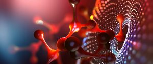
As an image can tell you more than 1000 words, imaging-based biomarker development is a powerful way to establish comparable studies between animals and humans ![]()

and animal level into humans, or vice versa.
Current and ongoing translational projects at the Centre, focusing on neurodegenerative diseases, cardiology and oncology, aim to characterise imaging-based biomarkers which can be identified and used in both animals and humans. These biomarkers can be defined as indicators which allow experts to differentiate between normal (healthy) or pathogenic biological processes, or to monitor pharmacologic responses to a therapeutic intervention. As an image can tell you more than 1000 words, imaging-based biomarker development is a powerful way to establish comparable studies between animals and humans.
Looking to the future
In the long term, the Centre will continue to promote synergies between different facilities to improve the translation of biomedical imaging at Ulm University and encourage new projects and collaborations. Other advantages of the Centre will be the standardisation of operating procedures and the provision of efficient scheduling, central data backup and archiving and unified finance management software. Furthermore, regular seminars and workshops are organised to increase awareness of the exciting opportunities that the Centre can provide, to offer chances to develop interdisciplinary research and to promote scientific exchange in the field of translational imaging. These regular seminars often include one of the Centre’s members presenting his/her imaging expertise. Future talks will especially target PhD students, to highlight the imaging techniques and core facilities that exist at the Centre and University and to also encourage them to consider integrating imaging techniques into their work.
The key to finding a therapy for a disease lies in the understanding of its basic cellular processes. Mutations or malfunctions on this level can be responsible for many malign changes in the cells, leading to severe diseases. By “just looking at the thing” as famous physicist R. Feynman said, many important results can be obtained from a molecular to a human level. Bringing experts from fundamental through to clinical imaging fields will certainly modify our view on biological processes and lead to a better understanding and, hopefully, to the treatment of the diseases of today.
You have initial funding for three years, where do you see the project going after this?
After the completion of our initial funding period, we will strive for long-term integration of the Centre into the academic structures of Ulm University to allow for continuous support of increasingly complex translational research projects.
What are the biggest hurdles you have had to overcome?
The establishment of a new academic centre involves many challenges. In our case, we had to make the centre and its services visible to a broad user group amongst the university. Having built up an online information platform, we presented our overall imaging infrastructure and emphasised the advantages of a centrally coordinated translational imaging facility to interested users. With regular seminars and workshops, we want to encourage researchers to broaden their horizons in other research fields and techniques, and to find a point of contact for new translational projects.
These kinds of centralised infrastructure centres will progressively be implemented in most of the research sites ![]()
Currently, the Centre is only focusing on linking different facilities at Ulm University and Medical Centre. Are you planning to collaborate with other Universities to share technologies and expertise?
The technologies and expertise of our core facilities are not restricted to users from Ulm University. Interested users from other research institutions and industries can easily request access to our infrastructure via the Centre for Translational Imaging platform. The key challenge is to promote our infrastructure nationally, for example, by using networks such as the German Research Foundations “RIsources” Portal, which contains information about scientific research infrastructures. We are also in close contact with several German Translational Imaging Facilities to exchange experiences and challenges.
The MoMAN centre is focused on one aspect of research, imaging, at one specific university. Do you think that these kinds of centres will become more common for other research techniques at other institutes?
We are absolutely certain that these kinds of centralised infrastructure centres will be progressively implemented in most of the research sites. The advantages are obvious: with continuously increasing costs and the huge variety of innovative research instruments available, the one-time acquisition of cutting-edge equipment made available to many researchers will be cost-effective and time-saving. Scientists can focus on their projects and make use of the services of the core facilities without dealing with instrument maintenance, time-consuming usage training and unreproducible results due to operating errors. Apart from imaging, Ulm University has already established further facilities, ranging from “omics” to bioinformatical services.
The Centre for Translational Imaging connects scientists and their techniques ranging from cellular imaging at a molecular level, via small animal imaging, to human imaging, or vice versa. The Centre serves as a central coordinating unit and structure with the capabilities to provide information to potential users as to which technique may be best suited to their research question. Their aim is to promote more scientific exchange between fundamental researchers and clinicians and to help foster translational projects.
Funding
German Research Foundation
Collaborators
The Centre for Translational Imaging MoMAN relies on the expertise of its research team at Ulm University and Medical Centre, consisting of:
- Prof A. Beer Department of Nuclear Medicine
- Prof M. Beer Department of Radiology
- Prof G. Grön Department of Psychiatry
- Prof A. Ludolph Department of Neurology
- Prof J. Kassubek Department of Neurology
- Prof J. Michaelis Institute of Biophysics
- Prof C. Gebhardt Institute of Biophysics
- Prof W. Rottbauer Department of Internal Medicine II)
- Prof V. Rasche Department of Internal Medicine II)
- Dr A. RückCore Facility for Confocal and Multiphoton Microscopy
- Prof P. Walther Central Facility for Electron Microscopy
- Prof T. Wirth Institute of Physiological Chemistry
Bio
 Julia Nagy studied Chemistry at the University of Munich and received her PhD in Biophysics from Ulm University. Since 2016, Julia has been the science manager of the Centre for Translational Imaging MoMAN at Ulm University.
Contact
Dr Julia Nagy
MoMAN – Center for Translational Imaging
Ulm University
Albert-Einstein-Allee 23
89081 Ulm, Germany
E: julia.nagy@uni-ulm.de
T: +49 731 500 33635
W: www.uni-ulm.de/en/einrichtungen/center-for-translational-imaging-moman/